Attention Deficit Neurophysiology and Smartphone Use
Attention Deficit Neurobiology and Digital Media Influences
Attention Deficit Neurobiology and Digital Media Influences
As a researcher intrigued by attentional disorders in modern contexts, I’ve unified studies on ADHD pathophysiology with explorations of digital media’s role in fostering similar traits. Starting with core ADHD neural markers, I extended to causal effects of smartphones and a rat model of overstimulation.
Core ADHD Pathophysiology: TMS-EEG and Neurocognition in Participants vs. Controls
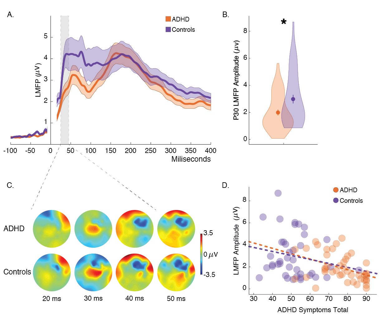
My foundational work examined right prefrontal cortex (rPFC) activity in ADHD. In this study, we compared 57 adults with ADHD to 54 matched controls using TMS-EEG over the rPFC and during a Stop Signal task (Hadas et al., 2021). ADHD participants showed reduced early TMS-evoked potentials (LMFP around P30, 25-45 ms post-TMS), correlating with symptom severity on the Conners’ Adult ADHD Rating Scale (CAARS) (rho = -0.37, p < 0.001).
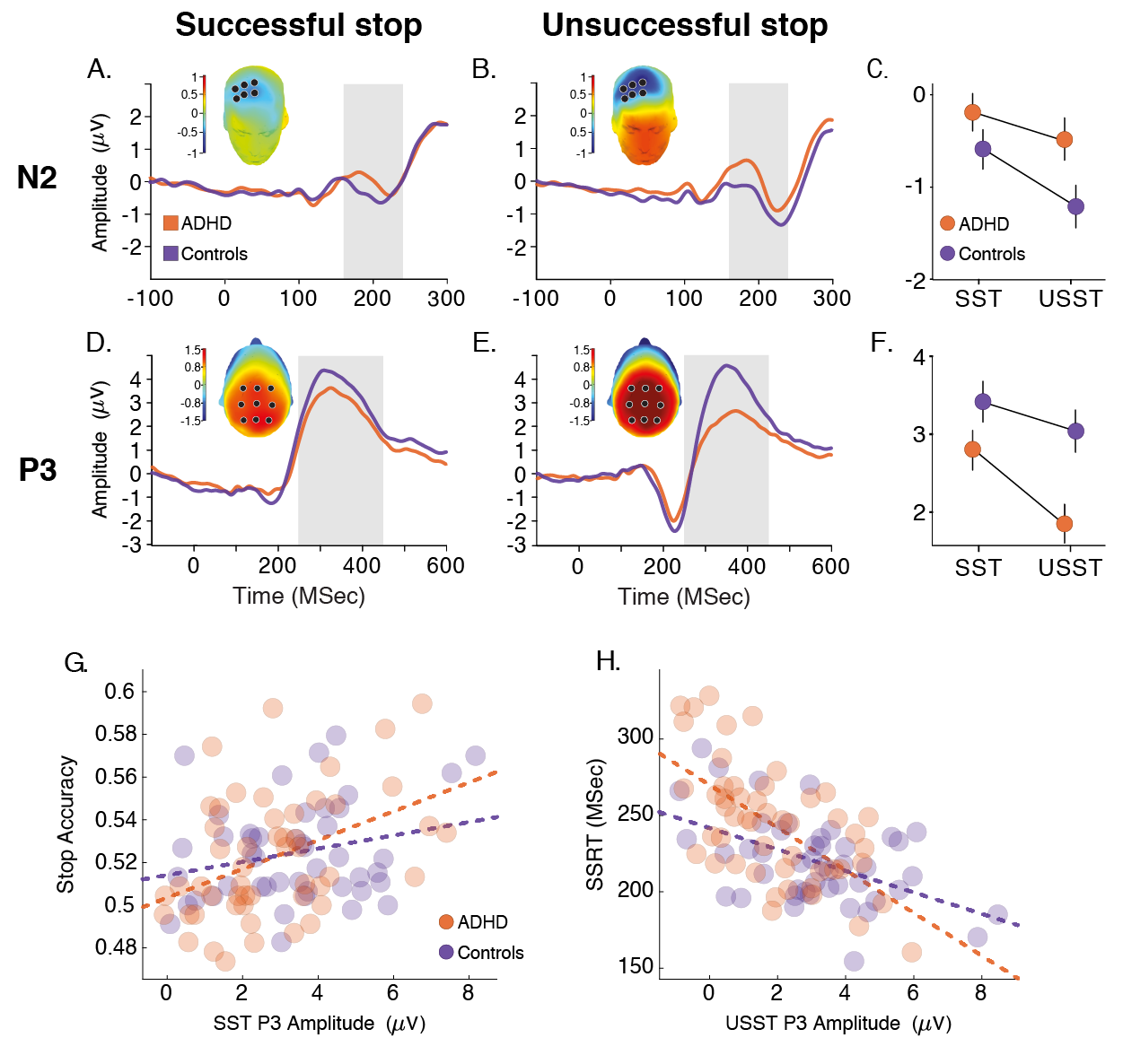
In the task, they had longer Stop Signal Reaction Times (SSRT) and higher errors, with diminished N2 (marginal, p=0.05) and P3 (p=0.012) ERP components—P3 linked to stopping accuracy (rho=0.4, p<0.004) and SSRT (rho=-0.59, p<0.00001).

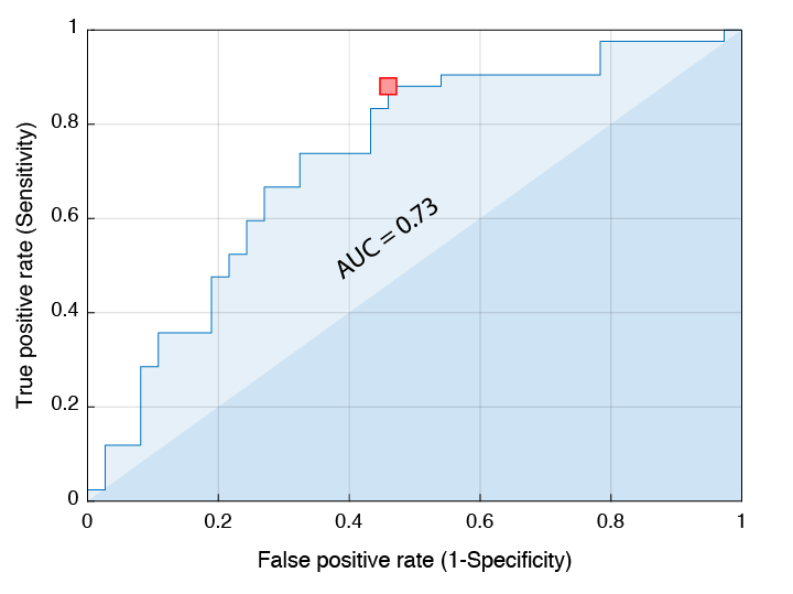
A linear discriminant model using these rPFC markers yielded 72% diagnostic accuracy (sensitivity 88%, specificity 54%). This established rPFC hypoactivity as a biomarker for ADHD severity and impulsivity, conceptually linking to how digital stimuli might exacerbate such deficits.
Digital Causality: Smartphone Exposure in Humans

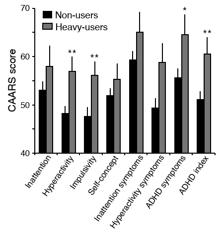
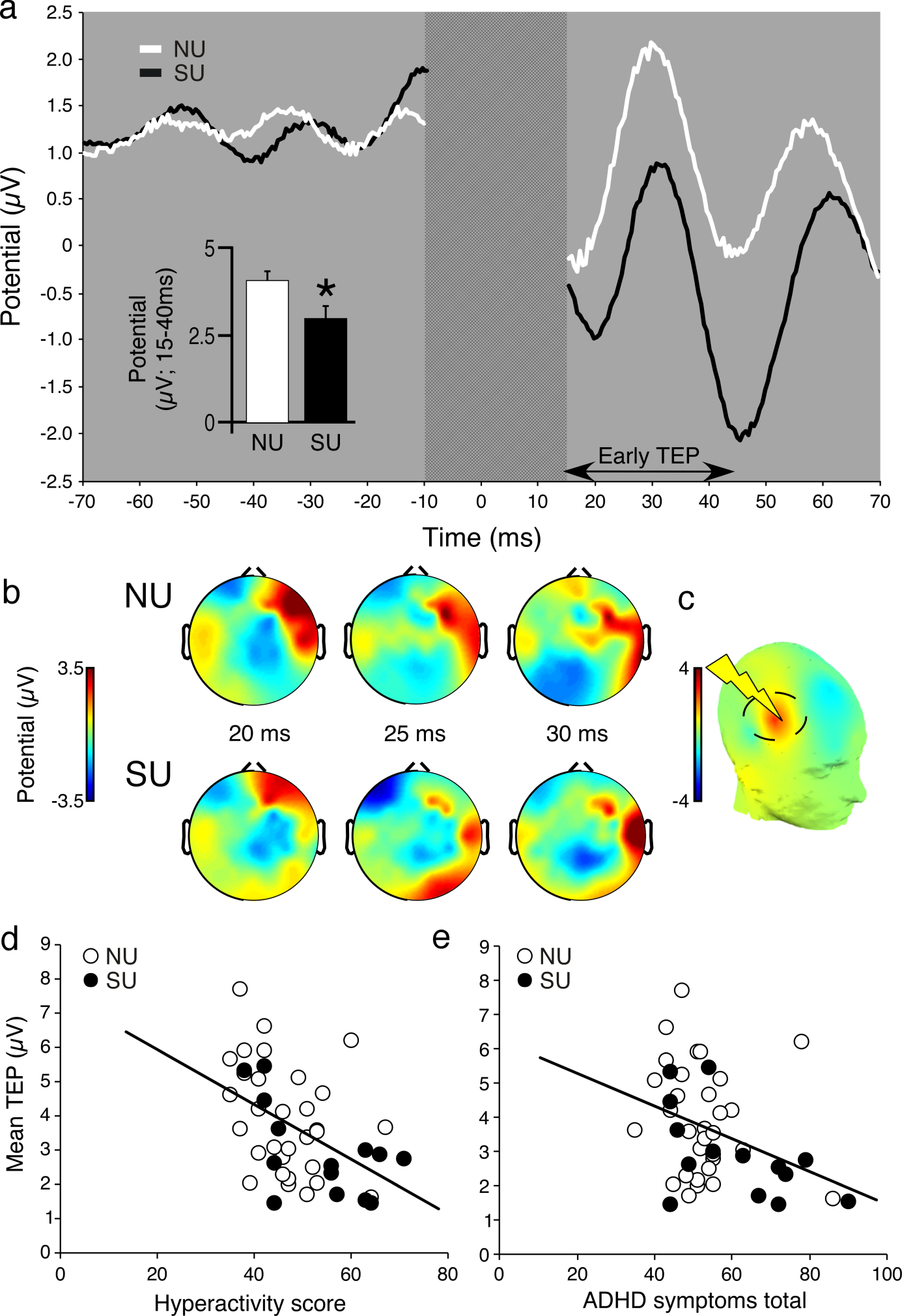
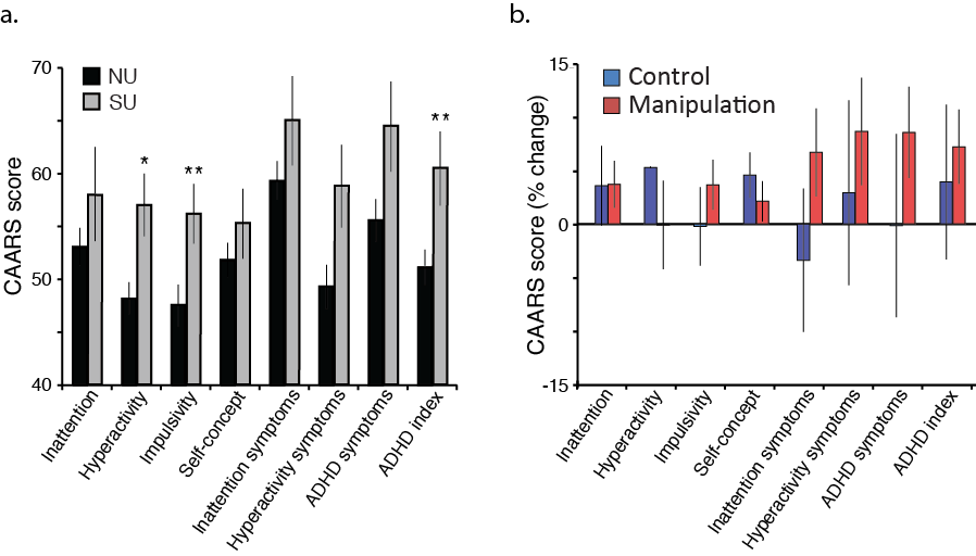
Inspired by ADHD patterns, we tested if heavy smartphone use induces similar traits, focusing on the right PFC—a key hub for impulse control, distinct from the left dorsolateral PFC (DLPFC) targeted in depression circuits. Comparing 16 heavy users to 35 non-users (basic phones), heavy users had elevated CAARS scores (inattention, impulsivity, hyperactivity) and Concern for Appropriateness Scale (CAS) values, plus steeper delay discounting and poorer numerical accuracy (Hadar et al., 2017).
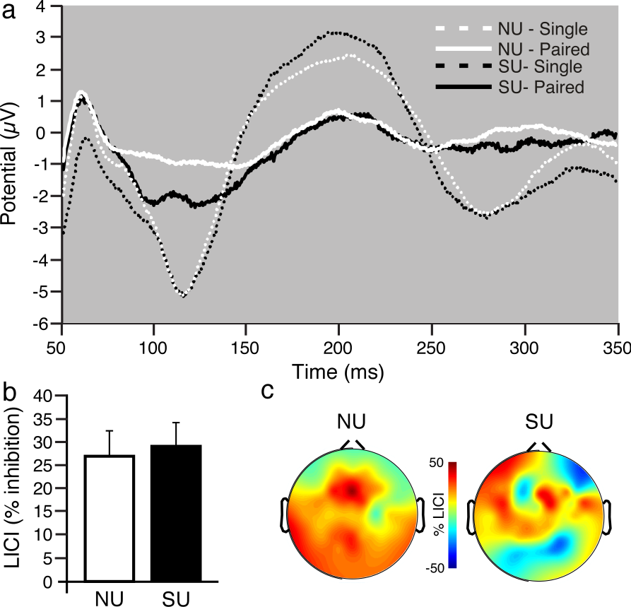
TMS-EEG over the right PFC revealed reduced early TEPs (15-40 ms post-TMS) and lower long-interval cortical inhibition (LICI), correlating with inattention severity (i.e., TEP rho = -0.45, p<0.01; LICI suppression reduced by ~20%).
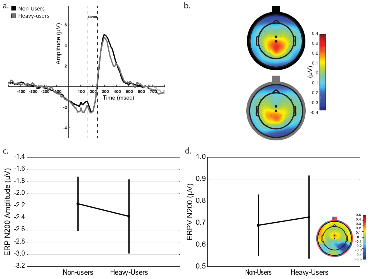
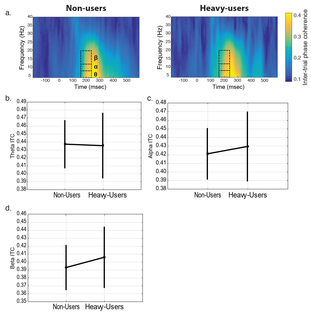
In the Stop Signal task, heavy users showed diminished N200 ERPs (p<0.05), reflecting impaired conflict monitoring, alongside reduced theta-band spectral perturbations and inter-trial coherence—EEG markers of attentional lapses tied to right PFC hypoexcitability.
Causally, we exposed 20 non-users (expanded from 12) to smartphones for up to six months (controls n=16). This increased inattention, CAS, and numerical deficits; usage predicted severity. Short-term right PFC changes were absent, implying gradual neural shifts
Mechanistic Model: Sensory Overload in Rats
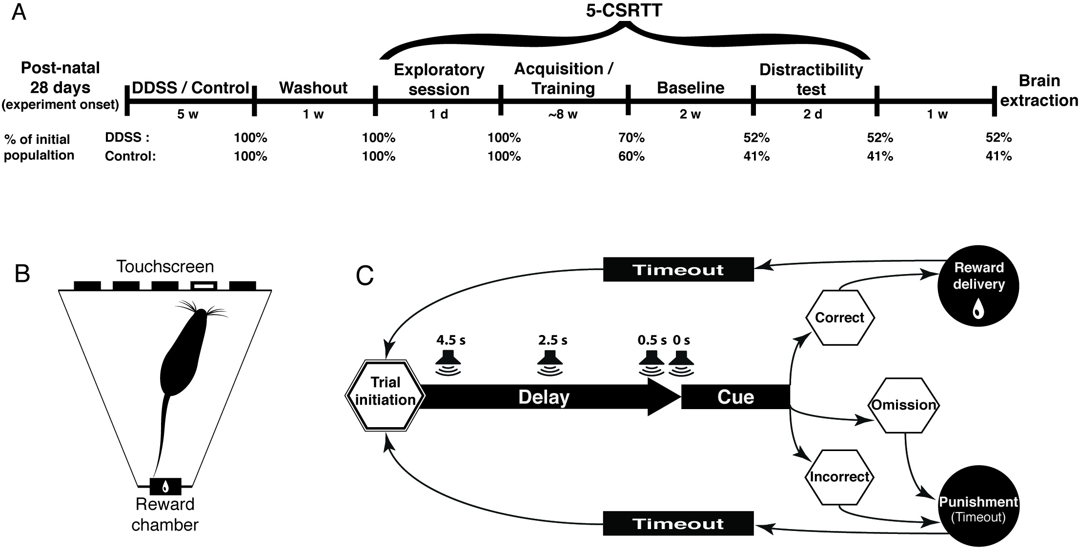
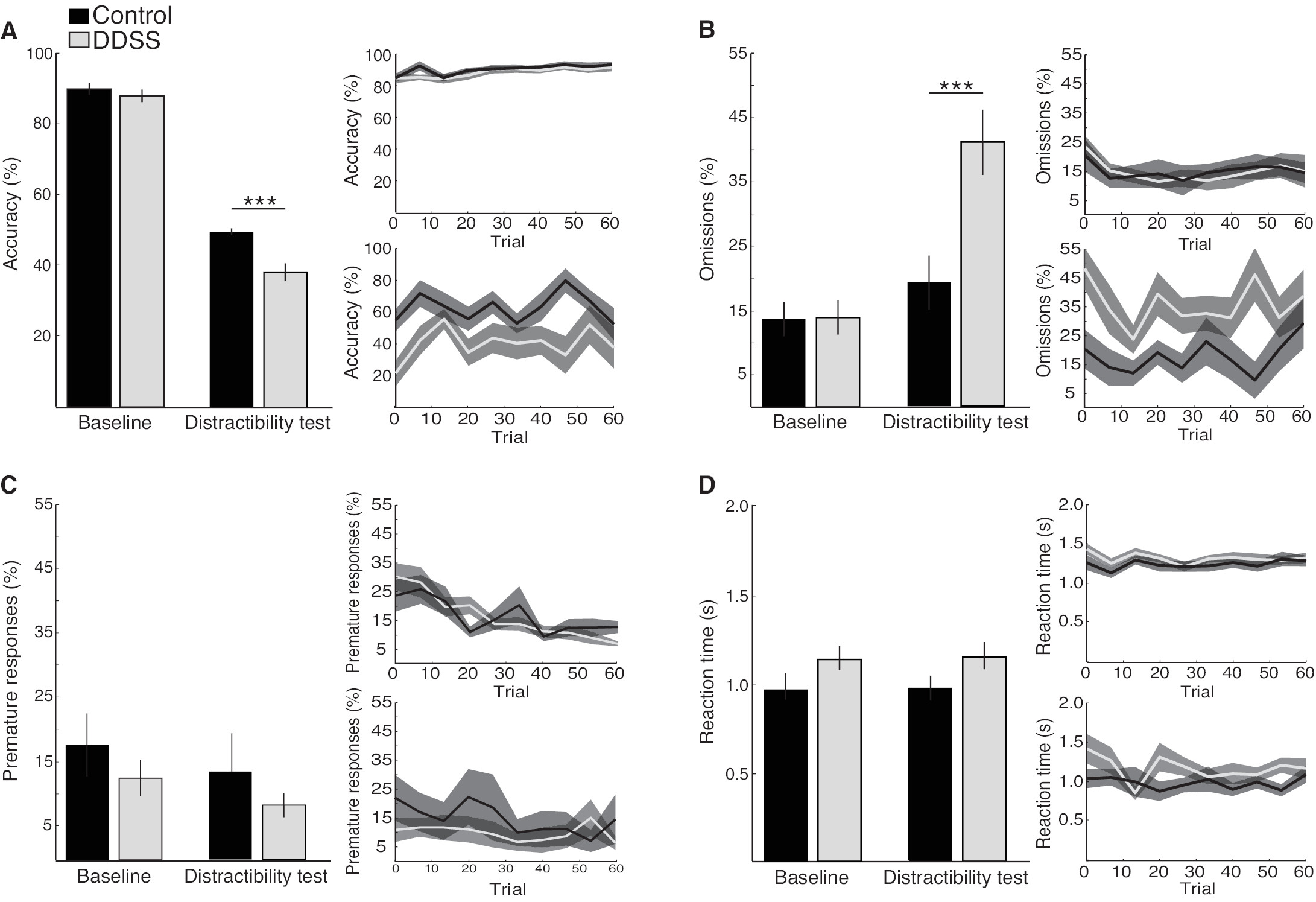
To probe biology, I developed a rat model: Juvenile rats received “developmental dynamic salient stimulation” (DDSS)—rapidly changing odors mimicking digital notifications—vs. static controls (Hadas et al., 2016) (Hadas, 2016). DDSS rats acquired the 5-Choice Serial Reaction Time Task faster but showed distractibility (more omissions, variable RTs under noise).
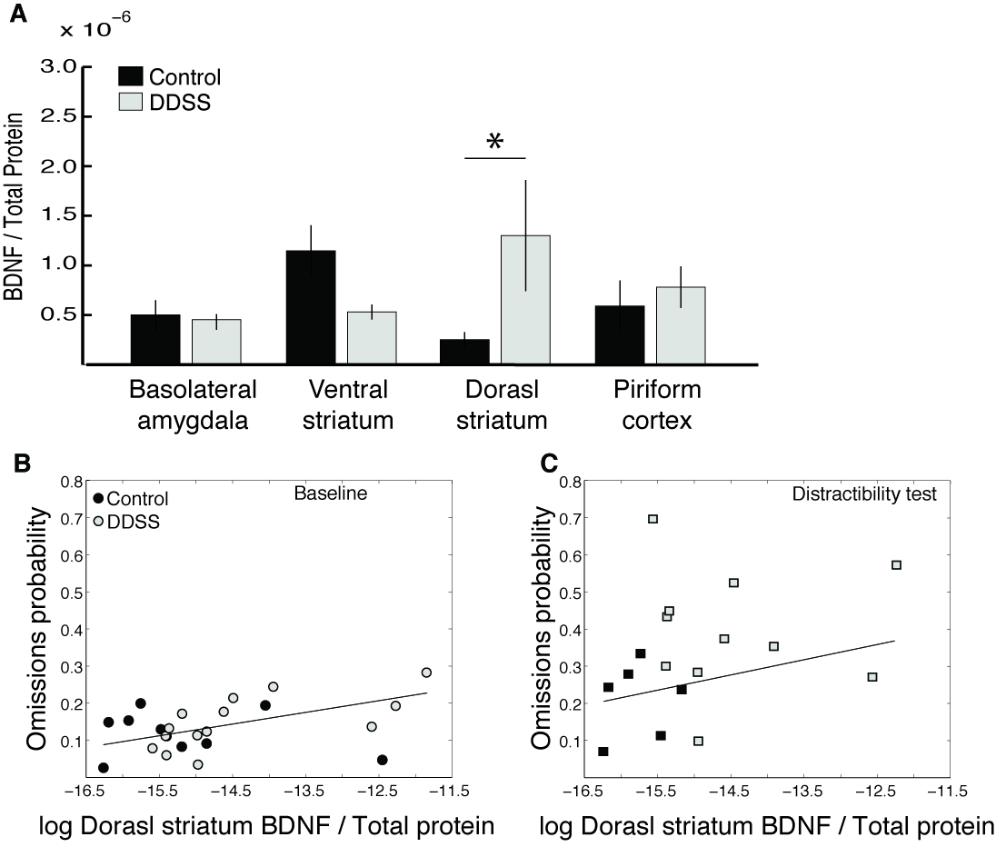
Dorsal striatal BDNF rose, correlating with errors, suggesting plasticity in reward salience.

Implications
This project ties rPFC hypoactivity in ADHD to digital-induced attentional erosion via striatal adaptations. It challenges heritability, highlighting environmental roles, and supports tech moderation.